

Raman Spectroscopy
The discovery of the Raman effect by C.V. Raman in 1928 marked a significant milestone in spectroscopy, earning him the Nobel Prize in Physics. Raman spectroscopy is a vibrational spectroscopic technique that probes molecular vibrations by analyzing the inelastic scattering of light, known as Raman scattering. This method has become indispensable in physical chemistry, materials science, and biophysics due to its ability to reveal detailed structural, electronic, and dynamic properties of materials without causing damage to the sample.
Raman spectroscopy's fundamental principle involves the interaction between monochromatic light and a sample. When light interacts with the sample, most photons scatter elastically at the same energy, a process known as Rayleigh scattering. However, a small fraction of the light undergoes inelastic scattering, resulting in an energy shift corresponding to the molecular vibrations within the material. These shifts create a unique spectral fingerprint that reflects the chemical composition and molecular structure of the sample.
Raman scattering consists of two distinct components: Stokes scattering, where the scattered light loses energy and shifts to longer wavelengths, and anti-Stokes scattering, where the scattered light gains energy and shifts to shorter wavelengths. Stokes scattering typically dominates because it arises from molecules initially in their ground vibrational state, while anti-Stokes scattering occurs less frequently and involves molecules in an excited vibrational state. The intensity ratio between Stokes and anti-Stokes scattering is temperature-dependent and offers valuable insight into the vibrational population and thermodynamic properties of the material.
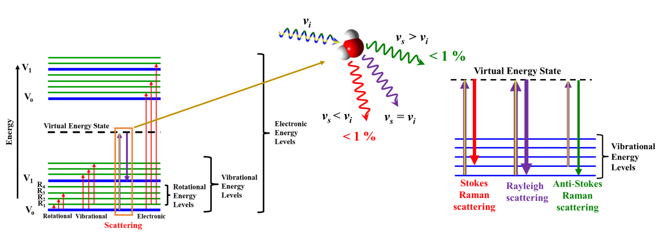
Figure 1: Illustration of a basic Raman spectroscopy setup and energy diagram of Raman and IR processes...
Limitations of Conventional Raman Spectroscopy
Despite its numerous advantages, conventional Raman spectroscopy faces several challenges. One major limitation is low sensitivity, as detecting molecules at very low concentrations is difficult due to the inherently weak Raman signal and the low probability of scattering events. Additionally, the spatial resolution of Raman imaging is constrained by the diffraction limit of light, restricting the ability to resolve features smaller than approximately half the wavelength of light (around 200 nm). This presents significant obstacles for nanoscale imaging. Fluorescence interference from certain samples can further overwhelm the weak Raman signal, complicating the acquisition of clear spectra.
To overcome these challenges, researchers have developed advanced techniques that leverage plasmonic effects and nonlinear optical processes to enhance Raman signals and broaden the scope of applications.
Plasmonic-Enhanced Raman Spectroscopy
Surface-Enhanced Raman Spectroscopy (SERS)
Surface-Enhanced Raman Spectroscopy (SERS) represents one of the most significant advancements in overcoming the limitations of conventional Raman techniques. SERS enhances sensitivity by utilizing the surface plasmon resonance (SPR) of nanostructured metal surfaces, such as gold or silver. When molecules are adsorbed onto or near these plasmonic surfaces, the localized electric fields amplify the Raman signal by several orders of magnitude. This enhancement enables the detection of trace amounts of analytes, making SERS particularly useful for monitoring molecular interactions and binding events in real-time.
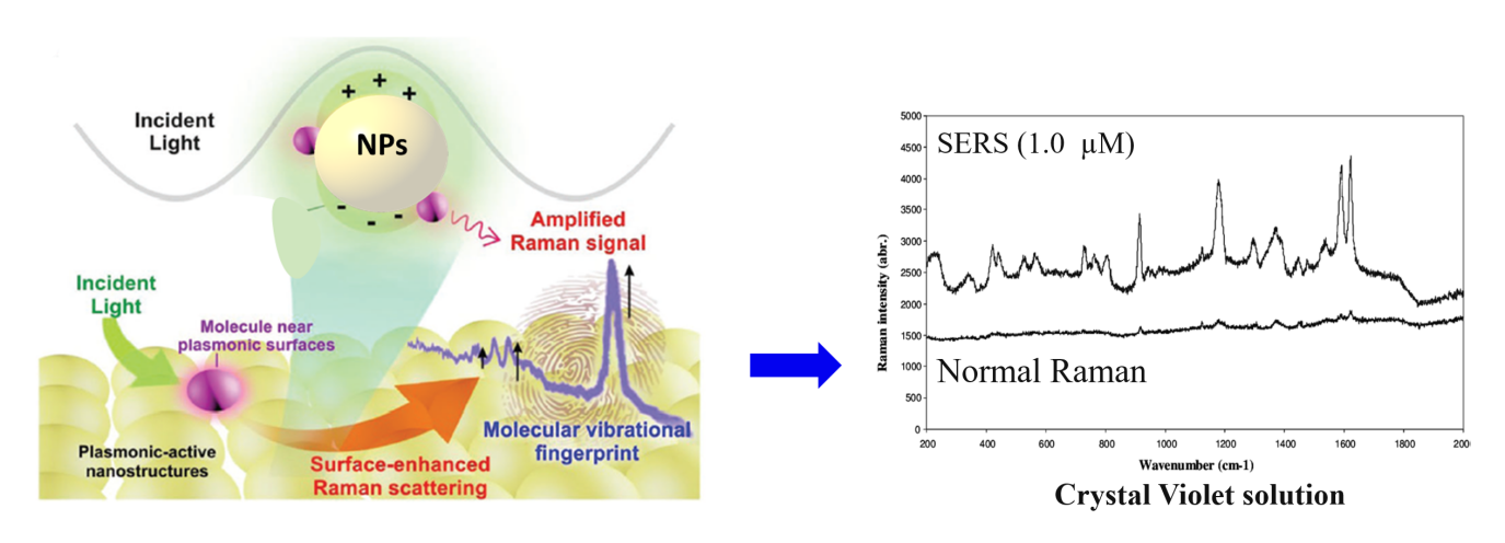
Figure 2: Schematic of plasmon resonance in SERS; showing the interaction between plasmonic nanoparticles and analyte molecules. SERS vs Raman spectrum
However, despite its potential, SERS faces several challenges, including reproducibility and uniformity in signal enhancement. Achieving consistent "hot spots"—regions where electromagnetic field enhancement is maximized—remains difficult, as variations in the morphology, size, and distribution of plasmonic nanostructures can lead to inconsistent signal intensities. Another frontier in SERS research involves expanding the range of plasmonic materials beyond conventional metals like gold and silver to include more sustainable, cost-effective, and functional materials, such as transition metals and hybrid nanostructures. Researchers are also striving to integrate SERS with advanced technologies like microfluidics, machine learning, and portable sensors to enhance real-time detection capabilities and field-deployable applications. Furthermore, improving the sensitivity of SERS for complex biological systems, where interference from non-specific binding or background signals can reduce accuracy, remains an active area of research.
Tip-Enhanced Raman Scattering (TERS)
Tip-Enhanced Raman Scattering (TERS) combines the principles of SERS with scanning probe microscopy (SPM), such as Atomic Force Microscopy (AFM) or Scanning Tunneling Microscopy (STM). In TERS, a sharp metallic or metal-coated tip, typically made of gold (Au) or silver (Ag), is brought to within a few nanometers of the sample surface. This sharp tip acts as a highly localized source of plasmonic enhancement, confining the electromagnetic field to a tiny region at the tip apex. The result is a dramatic amplification of the Raman signal, enabling the detection of individual molecules or nanoscale domains with sub-diffraction-limited spatial resolution, often down to the single-digit nanometer scale.
One of the key advantages of TERS is its capability to provide chemical imaging with both high spatial resolution and molecular specificity. This makes TERS particularly valuable for studying nanoscale features in materials science, surface chemistry, and biological systems. For instance, TERS can be used to analyze defects in graphene (hyperlink to my paper), molecular arrangements on functional surfaces, protein distributions on membranes, or the composition of biomolecular condensates.
Achieving optimal TERS performance depends on several factors, including the quality and sharpness of the tip, the plasmonic properties of the tip material, and the stability of the tip-sample interaction. One significant challenge is the fabrication of reproducible and durable tips that maintain their structural integrity over time and repeated use. Furthermore, TERS produces hyperspectral datasets that contain vast amounts of information, making data extraction and interpretation complex and time-consuming. Researchers are increasingly integrating TERS (or Raman imaging in general) with machine learning and multimodal imaging. These approaches aim to automate data analysis and enhance the interpretation of chemical imaging datasets.
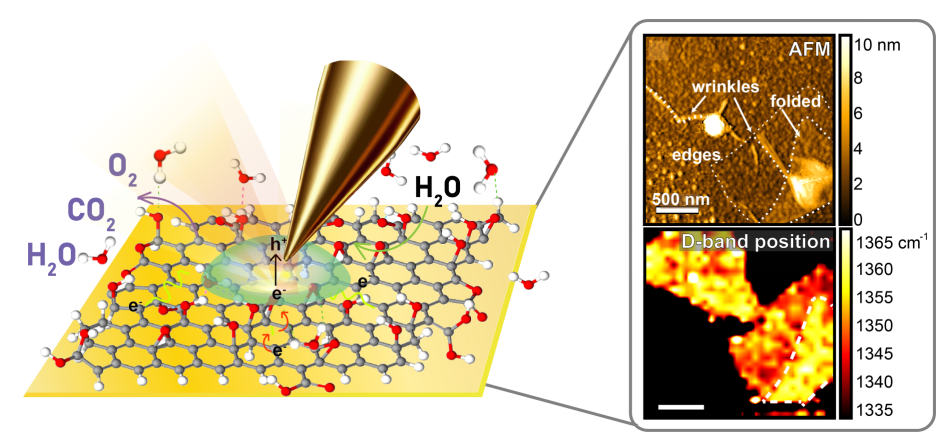
Figure 3: Diagram of TERS showing a plasmonic tip interacting with molecules on the substrate surface.
Nonlinear Optical Raman Techniques
Nonlinear optical Raman techniques, such as Stimulated Raman Scattering (SRS) and Coherent Anti-Stokes Raman Scattering (CARS), offer powerful solutions to enhance signal strength and suppress background interference, significantly expanding the applicability of Raman spectroscopy.
Stimulated Raman Scattering (SRS) amplifies Raman signals by utilizing an intense pump laser to drive stimulated emission, producing a stronger, highly detectable signal. Unlike spontaneous Raman scattering, which relies on rare inelastic photon interactions, SRS generates a signal only when the pump and Stokes beams interact with the sample, ensuring selective detection of vibrational modes. This process leads to a stronger and more detectable signal, drastically improving sensitivity and imaging speed. One of the key advantages of SRS is its ability to minimize background noise, as the signal occurs only when both the pump and Stokes beams are present, allowing selective detection of vibrational modes. This specificity makes SRS particularly useful for imaging live cells, tissues, and other biological samples without introducing photodamage. Because of its high sensitivity and ability to provide label-free imaging, SRS has become a powerful tool for tracking molecular interactions in real-time, revealing critical insights into dynamic biological processes.
Coherent Anti-Stokes Raman Scattering (CARS) complements SRS by addressing fluorescence interference through nonlinear optical processes. CARS generates anti-Stokes signals at higher frequencies than the excitation light, effectively enhancing the signal-to-noise ratio while suppressing background fluorescence. This allows for high-contrast imaging, particularly in biological samples with strong autofluorescence. CARS excels at real-time visualization of molecular structures and processes within living cells, offering a valuable tool for studying cellular dynamics and complex biological environments.
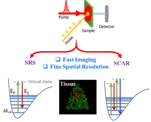
Figure 4: Energy diagram of CARS and SRS.
Summary of Techniques
Technique |
Enhancement Method |
Key Benefits |
Raman |
None |
Non-destructive, molecular fingerprinting |
SERS |
Surface plasmon resonance (SPR) |
High sensitivity, real-time molecular interactions |
TERS |
Plasmonic tip enhancement |
Improved spatial resolution, reproducibility |
CARS |
Nonlinear processes |
Fluorescence suppression, high signal-to-noise ratio |
This reference page is maintained by the You Lab at Westlake University. If citing, please reference: You Lab, Spatiotemporal Spectroscopic Imaging Laboratory, School of Engineering, Westlake University. youxiao@westlake.edu.cn